Core Imaging Facility
Core Imaging Facility
- Laser Scanning Confocal Microscopy: Zeiss LSM710
- Fluorescence and Transmitted Light Axio Imager A2 M
- Advanced Stereozoom Research Microscope - Stereo Discovery V20
- Polarized Light Microscopy - ZEISS AXIO Imager A2
- Student microscope- Primo Star HAL LED
- Student Stereomicroscope - Stemi DV4
1. Laser Scanning Confocal Microscopy: Zeiss LSM710
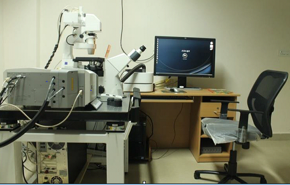
The Carl Zeiss Inc LSM710 has three fluorescence and one transmitted light detector. The LSM710 is particularly good for dim or photobleachable samples. Probes such as YFP and mCherry can be imaged using this instrument.
- Laser lines are 405, 458, 488, 514, 543, 594, 633
- Live Cell Imaging,
- Single Molecule Analysis,
- Imaging of Yeast
- The LSM 710 allows use of more dyes
- Low-noise electronics with up to 30% longer sampling time per pixel via oversampling
- Excellent contrast due to improved laser suppression by 100 to 1000 times (even with mirror-like samples)
- An increase in sensitivity due to a new spectral grating and spectral-recycling loop design (efficiency ≥ 90%)
- An array detector with three times lower dark noise
- Parallel 34-channel imaging over the entire wavelength range
- APD-imaging and photon counting
- Capable of continuous spectral detection over the whole wavelength range used simultaneously
- Multicolor imaging can be performed to perfection, allowing you to use the latest fluorescent proteins without spectral crosstalk
- The fast and flexible detection technology of the LSM 710 combined with the high performing In Tune (488 to 640 nm, > 1,5 mW per wavelength) means the fluorescence signal can be detected very close to the excitation wavelength. In addition, In Tune is the perfect flexible laser system for measuring fluorescence lifetimes of dyes (Pulse < 5 ps, 40 MHz) that couldn’t be examined before.
- It involves faster scan speeds at lower zoom factors (i.e., larger fields of view with a field number of up to 20 mm in the intermediate plane) or more consistent imaging conditions
2. AXIO Imager A2 M Fluorescence and Transmitted Light
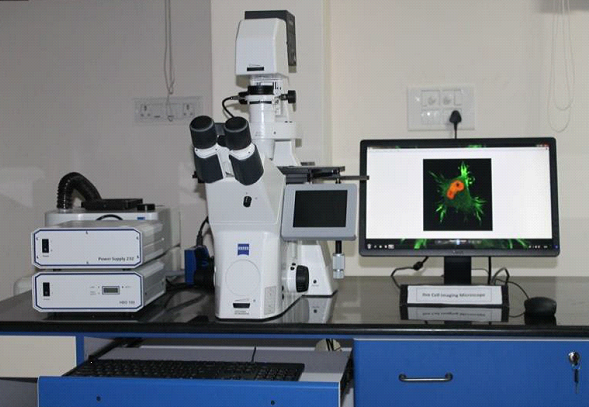
Upright research microscope for sophisticated imaging in fluorescence and transmitted light Brilliant optics and bright fluorescence. The integrated contrast manager and the programmable light manager guarantee defined conditions and reproducible results at all times.
Filters
- DAPI: Absorption maximum at a wavelength of 358 nm (ultraviolet) and its emission maximum is at 461 nm
- FITC: FITC has excitation and emission spectrum peak wavelengths of approximately 495 nm/519 nm
- Bright Field:Both reflected and transmitted properties available
3. Advanced Stereozoom Research Microscope - Stereo Discovery V20
Designed for Optimal Depth Perception and Maximum Zoom Range
Use the 20:1 zoom range to go from largest overview into the smallest details. All optical components deliver brilliant threedimensional images with exceptional depth perception. Precisely choose your optimal magnification or move to user-defined zoom positions with eZoom. Refind your magnification with an accuracy of more than 99 percent! The external touch panel SYCOP controls all essential functions of SteREO Discovery.V20: magnification, focus, contrast, brightness and more.
Highlights
- Motorized 20:1 zoom
- Seamless integration into the modular system of SteREO microscopes
- Up to 345x total magnification with 10x eyepieces
- Maximum resolution of up to 1000 LP/mm with PlanApo S 2.3x objective
- Motorized and encoded components for reproducible results
- Illumination and contrast methods based on cold light or LED
- Ergonomic and easy operation and control with the optional touch panel SYCOP
Precicion: eZoom Images – Twice as Sharp
The zoom body is the core of stereo and zoom microscopes. When zooming, lenses have to be positioned precisely. Until now, a metal component milled with great care would determine the exactness of this movement, and with it the optical quality of your microscope. eZoom replaces the mechanical curve with an electronic one. Stepping motors position the moveable lenses precisely and take the tolerances of the individual lenses into account. Each zoom body describes its own zoom curve and captures visibly more details. eZoom follows the base line for image sharpness over the magnification range with a doubled precision, compared to a mechanical zoom body. Program zoom curves individually.
When the micro clapper of the computer-controlled glue leveling machine brings eZoom’s lens in the zoom body into position, it is glued and cured with UV light.
The SYCOP Concept: Take Control
The SYCOP concept makes your work easier: use all essential functions of your stereomicroscope without moving your eye from the eyepiece. Control all components with joystick and touch screen or assign individual functions to the buttons. Position the SYCOP wherever you need it. Magnification and focus, control of brightness, display of current optical data - the control of your microscope has never been easier.
4. ZEISS AXIO Imager A2 Polarized Light Microscopy
Confidence thanks to automatic component identification
Microscope settings you can rely on at all times, on account of the Automatic Component Recognition ACR. All motorized stands recognize objectives automatically. Within the fully motorized Z-stand, ACR additionally identifies reflector modules. Exchange of components is registered by Axio Imager automatically.
Reliable long-term imaging thanks to vibration-free design
For time dependent measurements and high magnifications you can depend on the stability of Axio Imager. Nosepiece turret, z-guide and stage carrier have been designed as a compact, vibration-free unit, isolated from the rest of the stand. This "stable cell" creates ideal measurement conditions for superb results.
Comfortable operation due to extensive functionality
- Simplify complex procedures. Control of all motorized components is at your fingertips, by using the touchscreen at the stand or at the Docking Station.
- Save individual settings and retrieve them effortlessly at the push of a button. Operate the focus drive intuitively – thanks to ergonomically arranged control buttons with tactile surfaces.
- Alternatively operate the polarized light microscopy system via the freely positionable control panel, completely detached from the stand. The contrast- and light manager automatically selects the optimum settings in order to generate reproducible and reliable results.
Methods
Conoscopy: Fast and Reliable Crystal Analysis with Polarized Light Microscop
Capture orthoscopic and conoscopic image information simultaneously with Polarized Light Microscopy. With the specially designed Pol phototube, object, cross hairs and iris diaphragm are visible at the same time, thanks to an additional intermediate image plane. Thanks to the adjustable iris diaphragm this is also true for the limits of the conoscopic range down to a minimum crystal size of 10 µm. The pre-centered Bertrand optics are straightforward to turn on and off. This allows you to switch between techniques fast – even while capturing images or using a video device.
Consistent measurement performance
- Straightforward measurements usingthe rotary, ball bearing mounted stage with 360° scale and 0.1° vernier (e.g. for measuring cleavage angles in minerals)
- Determination of optical path differences or strain measurements
Compensators with fixed path difference
- Full -wave plate λ
- Quarter-wave plate λ / 4
- Full-wave plate λ, rotable +/- 8°
A wide spectrum of compensators is available, covering the measuring range from 0 to 30 λ.
Compensators with variable path difference
- Wedge compensator 0-4 λ
- Measuring compensators
- Berek tilting compensator 0-5 λ
- Berek tilting compensator 0-30 λ
Axio Imager is open to many more methods as for example:
- Thermomicroscopy and Digital analyses with Zeiss AxioVision software (e.g., with Grain Size Analysis or Particle Analyzer)
5. Student microscope Primo Star HAL LED
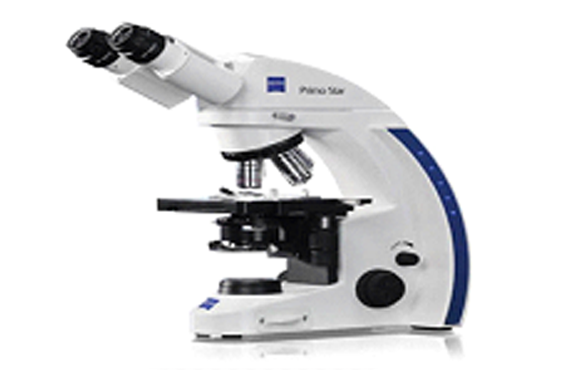
High performance for education and the Laboratory. Carl Zeiss Primo Star HAL LED Microscope is equiped with full-Köhler, stage R, Ph1, Ph2, Ph3 and FOV 20.
Description
Primo Star HAL microscope,full-Köhler, stage R, Ph1, Ph2, Ph3, FOV 20 Complete configuration with full-Köhler stand including:
- Halogen illumination with 6V 30W halogen lamp
- 4-position nosepiece, tilted backwards
- Mechanical stage 75x30, drive right and specimen holder with spring clip left
- Binocular tube 30°/20
- Eyepieces 10x/20 Br.foc.
- Objectives Plan-Achromat 10x Ph1, 20x Ph2, 40x Ph2 and 100x Oil Ph3
- Condenser 0.9/ 1.25 with H, Ph1, Ph2, Ph3 phase contrast slider
- Pinhole diaphragm, dia.: 30mm
- External power unit 100...240VAC/50...60Hz/30VA with country-specific adapters
- Dust cover
Color filter set blue, green, yellow
Interference filter green, d=45 mm for phase contrast
6V 30W halogen lamp (spare part)
6. Student Stereomicroscope - Stemi DV4
The innovative advances incorporated in this microscope are obvious not only in the color of the instruments and their compact, original design, but also, and most particularly, in their attractive price - all the result of state-of-the-art production technologies. Sturdy, hard-wearing technology, the very easy operation of all major function controls and no compromises whatsover in image definition and brilliance - that is the best way to describe this stereomicroscope from ZEISS. In addition to the four basic models, optionally with a zoom system or adjustable fixed magnifications, the compact Stand C and selected accessories (e.g. image documentation, darkfield, and many more) are other outstanding features.
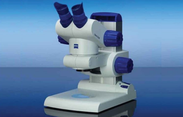
Biomedical Applications
- Hobby and private applications
Materials Applications
- Aerospace Industry
- Alignment Tasks
- Automobile Industry
- Building Materials Industry
- Dentistry
- Hobby and Private Applications
- Materials Technology
- Mechanical Engineering
- Numismatics
- Quality Inspection
- Semiconductor Technology
- Wood and Paper Industry