Scanning Electron Microscope
Scanning Electron Microscope
Scanning Electron Microscope
A Scanning Electron Microscope (SEM) is a type of electron microscope that produces images of a sample by scanning the surface with a focused beam of electrons. The electrons interact with atoms in the sample, producing various signals that contain information about the surface topography and composition of the sample.
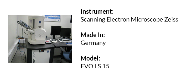
SmartSEM™ software, the EVO® LS 15 is a highly flexible, easy-to-use, high definition imaging and analysis tool delivering fast, accurate, repeatable results across all samples.
Applications
Scanning electron microscope is useful to study the morphology and investigate the mineralogy of nature resources, analysis of the minerals and rocks, conduct automated analysis of particles, analyze criminal evidence and toxicology. It can also be used to investigate and develop materials, analyze material properties.
Equipment Specifications
- Accelerating voltage - 2 to 30 kV
- Power Requirements- 100 - 240 V, 50 or 60 Hz single phase
- Magnification < 5 - 1,000,000 x
- Field of View - 6mm at the Analytical Working Distance (AWD)
- X-ray Analysis - 8.5mm AWD and 35° take-off angle
- Available Detectors BSD – Multisegment Diode
- ETSE – Everhart-Thornley Secondary Electron Detector
- VPSE – Variable Pressure Secondary Electron Detector
- SCD – Specimen Current Detector
- Chamber 365mm (Ø) x 275mm (h)
- OptiBeam®* Modes Resolution, Depth, Analysis, Field, Fisheye
- Pressure Range 10 - 400 Pa (MA Series)
- 10 - 3000 Pa (LS Series)
- Maximum Specimen Height 145 mm
Instruments Details: